As an English Setter of refined tastes and exacting standards, I often find myself observing my humans. They gaze into my soulful, deep-set eyes, whispering sweet nothings about how expressive they are. I appreciate the compliments, of course—high standards are meant to be acknowledged. But as I look back at them, I often wonder: do they actually know how much sophisticated machinery is working behind these gorgeous eyes of mine?
Today, we are going to talk about a piece of biological engineering that humans completely lack, yet we dogs possess in abundance: the third eyelid. Scientifically known as the nictitating membrane (or simply the “haw”), this hidden marvel is the unsung hero of canine vision. Let us dive into the fascinating, medically accurate, and altogether superior world of the canine eye.
What Exactly Is the Third Eyelid?
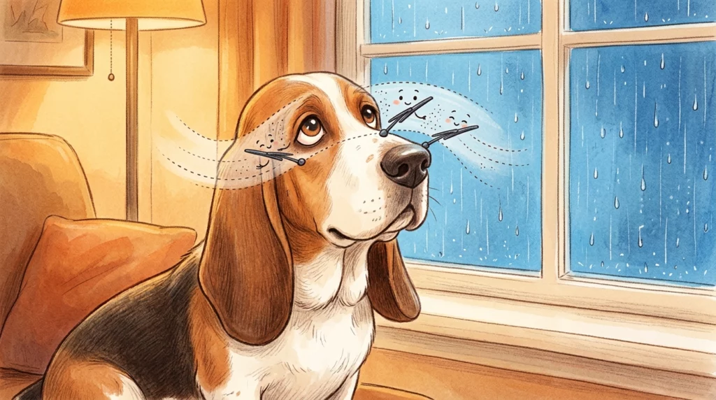
If you have ever caught your dog mid-nap, or perhaps looked closely at them while they were deeply relaxed, you might have noticed a pale pink or whitish tissue sliding horizontally across the corner of their eye. No, your dog is not secretly an alien. That is the third eyelid.
Unlike our upper and lower eyelids, which blink vertically, the third eyelid is positioned horizontally in the inner corner (the medial canthus) of each eye. As the experts at the Animal Vision Center of Virginia explain, this membrane is a specialized structure composed of a T-shaped cartilage frame, a highly active tear gland, and a protective layer of conjunctival tissue.
While humans are left to fend for themselves with just two eyelids—an evolutionary oversight I find deeply tragic—most mammals, birds, and reptiles boast this incredibly useful third layer of defense.
The Three Superpowers of the Nictitating Membrane
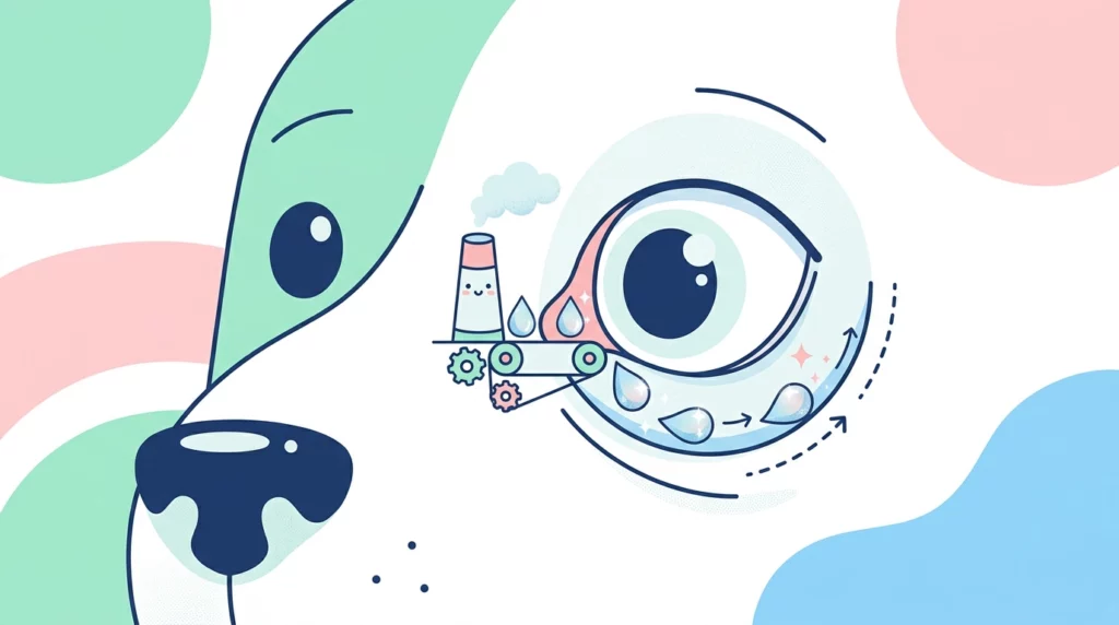
The third eyelid does not just sit there looking mysterious. It has a busy daily schedule keeping our eyes clean, moist, and healthy. In fact, it performs three vital functions that keep us looking and feeling our best:
1. The Ultimate Windshield Wiper
Have you ever run headfirst through a thicket of wild ferns, chasing the scent of a phantom squirrel? I do it regularly. It is marvelous, but it does leave one’s face covered in pollen, dust, and stray pine needles. This is where the third eyelid shines. When our eyeball retracts slightly into the socket, the third eyelid passively sweeps across the cornea like a highly efficient windshield wiper, clearing away debris before it can cause scratches or irritation.
2. The Canine Tear Factory
Our eyes require constant lubrication to remain healthy, and the third eyelid is a key player in this department. Tucked inside its base is an incredibly important lacrimal (tear) gland. According to the Schwarzman Animal Medical Center, the gland of the third eyelid is responsible for producing anywhere from 30% to 50% of the watery portion of a dog’s tear film. Without this gland, the eye simply cannot stay sufficiently moist.
3. An Immunological Shield
The third eyelid is also part of our body’s security system. It contains lymphoid tissue (lymph follicles) that acts as a local immune barrier. Think of it as a tiny, built-in security guard that identifies and fights off bacteria, viruses, and foreign invaders right at the entryway of the eye. For a more detailed look at this histology, the anatomical breakdowns at Veterian Key offer excellent insight into how these lymphoid structures function.
The Vanishing Act: Why You Rarely See It
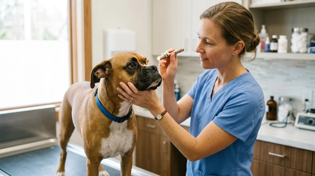
You might wonder why, if this membrane is so busy, you don’t see it constantly blocking our vision. The truth lies in its clever mechanical design.
Unlike birds or reptiles, who can voluntarily blink their third eyelids to protect their eyes while flying or hunting, dogs do not have direct muscular control over this membrane. Instead, it operates passively. When we are awake and alert, our extraocular muscles pull our eyeballs forward, keeping the third eyelid tucked neatly out of sight in the corner of the eye.
However, when we sleep, relax deeply, or feel unwell, the muscles around the eye relax, allowing the eyeball to sink slightly back into the socket. This physical shift pushes the fat pad behind the eye, which in turn pushes the third eyelid up and across the cornea. As noted by the veterinary resources at ToeGrips, this is why you will most often catch a glimpse of the “haw” when your dog is happily snoozing on the rug or just waking up from a serious nap.
When the Magic Glitches: What to Watch Out For
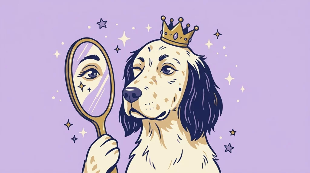
As a discerning dog, I must stress that while the third eyelid is a masterpiece of biology, it can sometimes experience health issues that require your immediate attention. Here are the two most common reasons the third eyelid might make an unwanted, permanent appearance:
“Cherry Eye” (Prolapsed Gland)
This is perhaps the most well-known issue involving the third eyelid. It occurs when the fibrous attachment holding the tear gland in place weakens or tears, causing the gland to pop out of its normal position. When this happens, a bright pink or red, cherry-like mass appears in the inner corner of the dog’s eye.
While it looks quite dramatic and alarming, it is usually not intensely painful in its early stages. However, as the experts at VCA Animal Hospitals emphasize, it is crucial to seek veterinary care quickly. If left untreated, the exposed gland can become inflamed, swollen, and infected.
Historically, vets would simply remove the protruding gland. However, because this gland produces up to half of our tears, removing it often leads to a lifetime of Dry Eye, officially known as Keratoconjunctivitis Sicca (KCS). Today, modern veterinary medicine favors surgical repositioning—tucking the gland safely back where it belongs. You can read more about these advanced surgical options on Veterinary Partner.
A Permanently Visible Third Eyelid (Elevated Haw)
If you notice that your dog’s third eyelid is staying up and visible even when they are fully awake and active, this is a clear signal that something is amiss. It can be a sign of:
- Dehydration or weight loss: When a dog is dehydrated or loses a significant amount of weight, the fat pad behind the eye shrinks, causing the eyeball to sink and the third eyelid to rise.
- Ocular pain: If the eye is injured or scratched, the dog will instinctively pull the eyeball back for safety, raising the membrane.
- Neurological conditions: Conditions like Horner’s Syndrome can affect the nerves supplying the eye, causing the third eyelid to droop or elevate abnormally. Detailed clinical notes on these neurological pathways can be explored via Veterinary Information Network (VIN).
If you ever spot a persistent change in how your dog’s third eyelid looks, do not guess. Take them to the veterinarian for a proper examination. Our eyes are precious cargo!
Niki’s Final Verdict
So, the next time you look into your dog’s beautiful eyes, take a moment to appreciate the incredible, invisible protective shield that keeps them sparkling. The third eyelid is just one of many reasons why we dogs are, quite frankly, marvels of design. Now, if you will excuse me, my third eyelids are calling for a very thorough, highly restorative afternoon nap on the sofa. Keep those standards high, dear friends!